
Journey to the ICU- Insane in the Drain!
- Miki Shibata

- Aug 18, 2019
- 5 min read
Author: Miki Shibata, MS, CCC-SLP Edited by: Ainsley Martin, MS, CCC-SLP
In this installment, we’ll go over some of the more common drains that a clinician may encounter in the ICU as well as precautions/considerations for each one. Drains are placed to remove bodily fluids whether for waste management, post surgery, and/or to regulate pressure.
Pigtail Catheter and Chest Tube


Pigtail catheters are small tubes placed in the abdominal or thoracic cavity to drain fluids blood, and/or air. Chest tubes are inserted into the pleural space (the space between the inner lining and the outer lining of the lungs) and serve a similar function to the pigtail catheters- helping to remove fluids, blood, and/or air from around your lungs, esophagus, or heart- to allow the lungs to fully expand. Both the pigtail catheter and the chest tube connect to a drain.
Indications for use:
- Trauma to the chest
- After surgery in chest
- Pneumothorax (commonly known as collapsed lung, air in pleural space)
- Hemothorax (bleeding in pleural space/ cavity)
- Drainage of pleural effusions (build of body fluids in the chest) often due to bleeding into the chest, fatty fluid collection, abscess in the lung or the chest (i.e. empyema), or heart failure (transudative pleural effusion - fluid leaking into pleural space due to increased pressure in the blood vessels or a low blood protein count)
Precautions:
- When positioning patients be mindful of location of chest tubes/pigtail catheters and make sure to have enough slack to complete the positioning change
- Drains must be kept upright and in "dependent position" meaning below the level of placement ...otherwise they won't drain.
- If tube is accidentally removed, place clean linen/gauze over site and apply pressure. Alert RN.
- If pt is experiencing sudden respiratory difficulties or increased pain, alert RN.
Closed Suction Drain with Bulb

Commonly known by the commercial name Jackson-Pratt drain or JP drain, this is a drain that is made of a small tube and bulb like collection unit. When the bulb is compressed, the drain uses negative pressure for removal of blood and fluids from insertion site. Once the drain is halfway full, usually the fluid is drained from the bulb, output quantity is measured, and sometimes the fluid is cultured.
Indications for use:
- Drain bodily fluids after surgery
Precautions:
- If the tube dislodges, apply pressure to the site with clean linen or gauze and alert the RN.
- Bulb should be kept in dependent (below the insertion site)
Hemovac
Indications for use: Just like the JP drain, the hemovac uses negative pressure when compressed to remove blood and fluid from the insertion site. This is normally utilized following orthopedic procedures.
Precautions: - If the tube dislodges, apply pressure to the site with clean linen or gauze and alert the RN.
- Bulb should be kept in dependent (below the insertion site)
Urinary Catheter

Two types of catheters are frequently seen in the hospitals, indwelling and external catheters (e.g., condom catheter, Purewick catheter).
Indwelling Catheters
Also known as Foley Catheter or a Foley, this indwelling catheter is a flexible tube that is inserted into the urethra of a patient who cannot urinate on their own. The catheter has a balloon at the end that is inflated to keep it in place. Urine output is measured to help determine adequate kidney function.
Indications for use:
- Urinary incontinence (leaking urine or not being able to control urination)
- Urinary retention (not being able to urinate, and retaining urine in the bladder)
- Surgery on the genitals or prostate
Precautions:
- Urine bag must be kept in dependent position to prevent old urine from entering back into the catheter.
- Move the urine bag as necessary during re-positioning to prevent pulling the line.
- Purewick devices are usually connected to suction cups. If you need to use oral suction you can grab a new suction cup/yankauer/tubing and connect it using a bifurcated suction regulator or simply disconnecting the suction cup for the Purewick and connecting the oral suction line to the wall. However, always ask the RN for permission and when reattaching be mindful that Purewick suction rate is usually a lot lower than for oral suctioning, so check with RNs regarding the proper suction rate.
Vacuum Assisted Closure System or Negative Pressure Wound Therapy
"Negative Pressure Wound Therapy" attributed to https://vimeo.com/leehealth
If you're fortunate enough to work at a polytrauma/burn center or a hospital that has a robust reconstructive surgery program, you might encounter one of these vacuum dressing devices. A sterile, open-cell foam is packed into the wound (to as a filter, preventing clogging from material such blood clots or dead tissue), covered with an occlusive dressing, and a pump that is programmed at a preset suction strength (usually between -125 to -75 mmHg )/rate (continuous/intermittent) is connected. This allows for drainage and moisture pulling from the wound site.
Indications for use:
- Ulcers (diabetic, venous, arterial, pressure)
- Burns
- Chronic wounds with large amounts of drainage
- Surgical/acute would at high risk for infection
Precautions:
- Some patients can be temporarily disconnected from the pump for mobilization. However, a MD order is required.
- Make sure to protect the occlusive dressing/foam as disruption can be very painful for the patient as well as problematic in wound management.
- If the negative pressure environment is disrupted, apply sterile dressing and report to RN for further wound management.
Intraventricular Catheter System (IVC), External Ventricular Drain (EVD), or Ventriculostomy
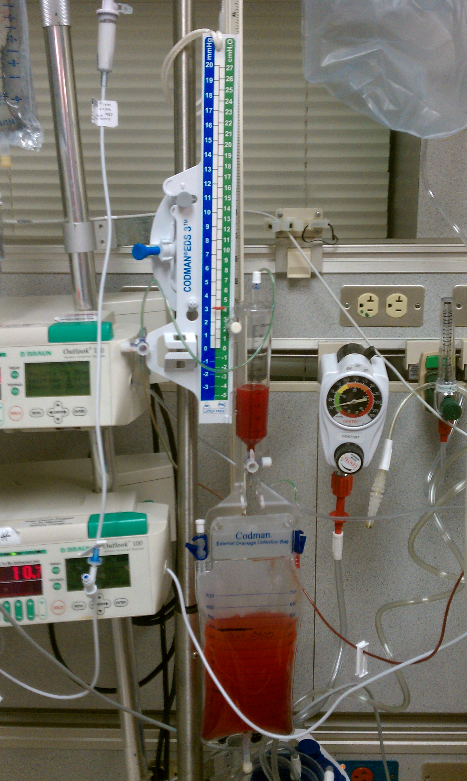
* Please also image search "EVD" for more pictures of what the attachment site looks like!
An EVD is often seen in the Neuro ICU. It's placed to remove blood product and cerebral spinal fluid (CSF) as well as to monitor intracranial pressure (ICP). This drainage system works based on pressure gradient vs. based on gravity like some other drains.
Fun fact! In over 90% of cases, preferred insertion site of the intraventricular catheter is the right cerebral frontal hemisphere for it's non-dominance for language.
Indications for use:
*Usually placed when ICP is above 20 mmHg. The range for normal adults ICP is 0 -15 mmHg.
- Intracranial hemorrhage with intraventricular extension
- Subarachnoid hemorrhage
- Traumatic brain injury
- Bacterial meningitis
Precautions:
- This drain is very sensitive to height, so don't elevate the head of bed or the height of the bed before checking with the RN.
Lumbar Drain
*Please image search "lumbar drain" to see what it looks like! Unfortunately, we were unable to find images with a copyright that allowed sharing!
A small, flexible plastic tube placed in lumbar subarachnoid space (L2-L3, L3-L4, or L4-L5) to remove cerebral spinal fluid (CSF). The drain is placed for collection of CSF after brain/spine surgery or to reduce pressure around the spinal cord or brain. It is also used to assess whether a shunt is needed when treating normal pressure hydrocephalus.
Indications for use:
- After thoracic aortic aneurysm dissection and repair
- Normal pressure hydrocephalus
- Cerebral spinal fluid leak
Precautions:
- Often patients with a lumbar drain have strict positioning requirements (most are head of bed flat). Check with RN and MD orders before attempting any head of bed elevation.
After reading this post, we hope you have a greater knowledge of various drains you may encounter in the ICU. If you have not yet had the opportunity, be sure to check out one of our previous articles on different lines you may encounter. As always, stay tuned for our next installment in the Journey to the ICU series!
References:
Lynn, S. J. (2017, June 20). Caring for patients with lumbar drains. Retrieved from https://www.americannursetoday.com/caring-patients-lumbar-drains/
Mancini, M. C. (2019, April 3). Chest tube insertion: MedlinePlus Medical Encyclopedia. Retrieved from https://medlineplus.gov/ency/article/002947.htm -- (Mancini, 2019)
Muralidharan R. (2015). External ventricular drains: Management and complications. Surgical neurology international, 6(Suppl 6), S271–S274. doi:10.4103/2152-7806.157620
Wechter, D. G. (2018, January 7). Closed suction drain with bulb: MedlinePlus Medical Encyclopedia. Retrieved from https://medlineplus.gov/ency/patientinstructions/000039.htm --(Wechter, 2018)
Wechter, D. G. (2018, January 7). Hemovac drain: MedlinePlus Medical Encyclopedia. Retrieved from https://medlineplus.gov/ency/patientinstructions/000038.htm -- (Wechter, 2018)


Comments